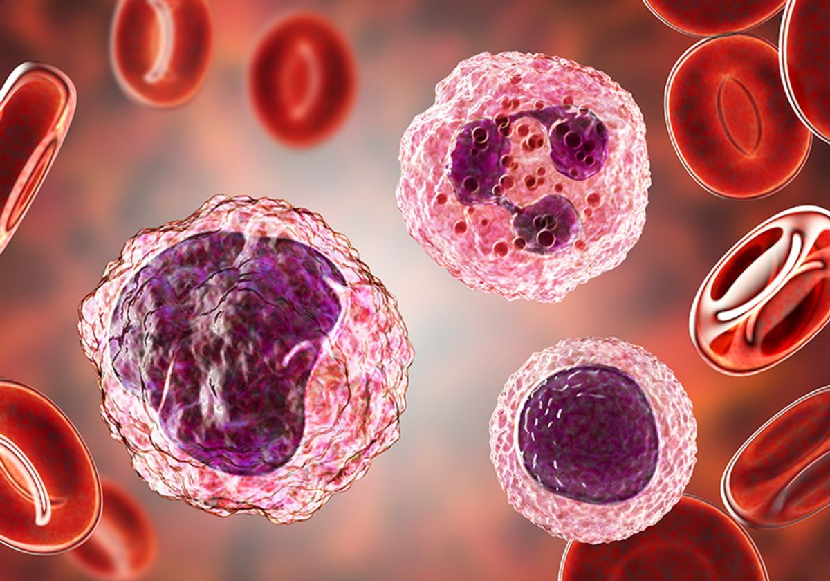
Cells are the basic building blocks of organisms and their morphology is dependent on their function, health, differentiation, and even response to treatment.1 Consequently, cell morphology serves as a visual indicator of the molecular and physiological processes that are occurring within the cell during normal and pathological conditions.2–4 Given the importance of cell morphology, scientists have now defined a new -omics category called morpholomics.
Drawbacks of Existing Morphology-Based Analytical Techniques
Researchers have used two primary methods to assess cell morphology: microscopy and flow cytometry. Microscopic examination mostly relies on image interpretation, where the human eye analyzes cell shape, roundness, and pigmentation. Moreover, fluorescent antibodies, dyes, and tagged biomarkers can label cellular features, such as lamellipodia or immune receptors, to help identify cell populations. However, microscopy-based analysis is a slow process, and it is hard to objectively quantify subtle morphological differences between cells.
Through flow cytometry, scientists analyze morphological characteristics, such as cell size and internal complexity, known as granularity. They also identify and quantitate cell populations through the absence or presence of specific biomarkers, where they can simultaneously label multiple cellular features with different fluorophores to provide multidimensional morphological information about their samples. Yet, flow cytometry only permits researchers to examine labeled cells and ignores unlabeled cell types which could be relevant to the disease or process they are studying.
Scientists often need to examine specific cell populations through downstream applications, such as sequencing or proteome analysis. However, subpopulations within heterogeneous cell samples may not occur at high enough frequency for further analysis and, thus, require enrichment. Researchers employ fluorescence-activated cell sorting (FACS), which is a flow cytometry-based technique, to isolate fluorescently labeled cells, but this method does not provide them with unlabeled cells for additional experiments. Conversely, they can identify unlabeled cell populations through microscopy, but enrichment is not attainable.
In-Depth Morphological Analysis and Gentle Population Isolation
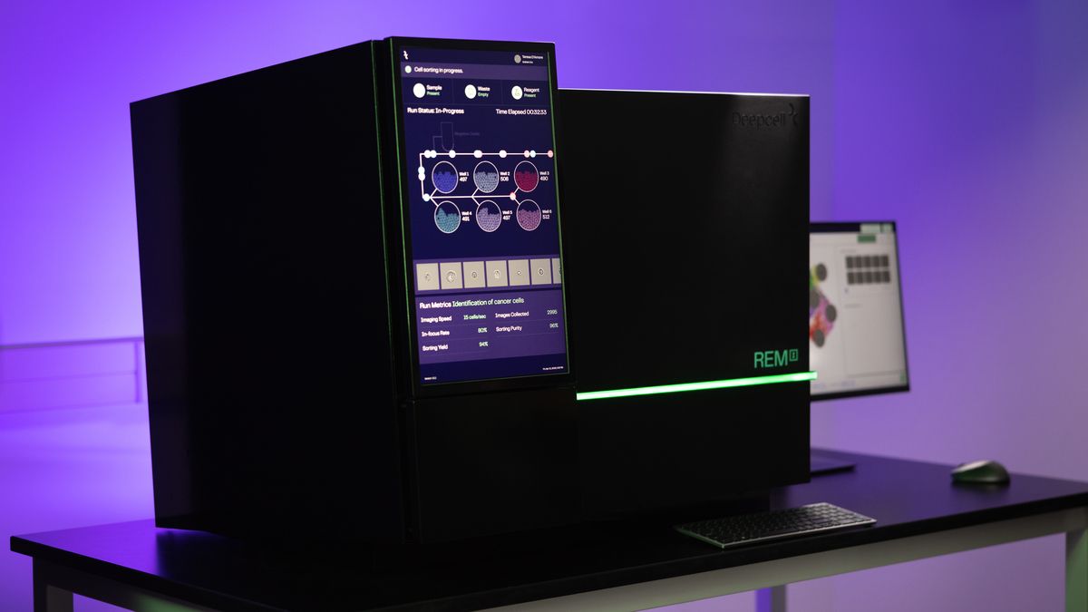
A new technology combines morphological analysis with unlabeled cell sorting and enrichment. The REM-I™ is a microscopy-based benchtop instrument from Deepcell™ that uses artificial intelligence (AI) to analyze cell morphology from high-resolution brightfield images. The instrument individually images unlabeled human cells while they are gently flowing through a microfluidic chip. Unlike morphological analysis by eye, which can miss a great deal of information, the platform’s AI model, called the Human Foundation Model (HFM), accurately and efficiently extracts these phenotypic details from each cell. As the HFM was pretrained by observing millions of cell images, the REM-I platform does not require additional training, so it is broadly applicable to a wide range of research areas, including cancer, immunology, cell and gene therapy, and drug screening.
The REM-I’s accompanying software, the Axon data suite, groups together cells that share the same morphological features and color codes these clusters, permitting the population’s further analysis. Additionally, the platform enables the gentle sorting of Axon-identified cell clusters into the microchip’s six wells. Unlike FACS, this user-chosen sorting is only dependent on cell morphology and does not require fluorescent labels, which ensures that the cells are viable and ready for functional and molecular analysis.
Morpholomics in Action
Recently, scientists from Deepcell and the University of California, Los Angeles used the REM-I platform to study and isolate carcinoma cells from patient effusion samples.5 Normally, these cells are not abundant in effusion samples, which prevents their further molecular characterization. However, with the REM-I platform, the researchers detected and enriched for the carcinoma cells, which allowed them to perform whole genome and targeted mutation sequencing on this population. Results like these highlight the platform’s functionality and the significance of morpholomics.
References
- Prasad A, Alizadeh E. Cell form and function: Interpreting and controlling the shape of adherent cells. Trends Biotechnol. 2019;37(4):347-357.
- Bakal C, et al. Quantitative morphological signatures define local signaling networks regulating cell morphology. Science. 2007;316(5832):1753-1756.
- Wu PH, et al. Evolution of cellular morpho-phenotypes in cancer metastasis. Sci Rep. 2015;5(1):18437.
- Yin Z, et al. A screen for morphological complexity identifies regulators of switch-like transitions between discrete cell shapes. Nat Cell Biol. 2013;15(7):860-871.
- Mavropoulos A, et al. Artificial intelligence-driven morphology-based enrichment of malignant cells from body fluid. Mod Pathol. 2023;36(8).


