Login
Subscribecell biology
Capturing the Brain Tumor Microenvironment with Tissue Engineering
Deanna MacNeil, PhD | Aug 4, 2023 | 3 min read
Researchers built a 3D glioblastoma model to study therapeutic resistance and improve drug screening systems.
Cryo-EM: Building on a History of Invention and Innovation
Thermo Fisher Scientific | Aug 2, 2023 | 1 min read
From humble yet ingenious beginnings to Nobel recognition, cryogenic electron microscopy (cryo-EM) provides insights into scientific questions that other technologies are unable to answer.
Enhancing Cell Morphology-Based Analysis
The Scientist’s Creative Services Team and Deepcell | 3 min read
Learn how the latest AI-driven technology uses morphology to comprehensively analyze and sort cell populations.
Inducing Cardiomyocyte Maturation
Jennifer Zieba, PhD | Aug 1, 2023 | 2 min read
By combining calcium and electrical pacing, researchers designed a scalable protocol for culturing mature cardiac tissues from induced pluripotent stem cells.
Captivated by the Great Expanse of Neurons
Danielle Gerhard, PhD | Aug 1, 2023 | 2 min read
According to Erin Schuman, science driven by fascination rather than tools will guide new discoveries.
A Novel Stem-Cell Derived In Vitro Model of Intestinal Inflammation
The Scientist’s Creative Services Team | 1 min read
In this webinar, Bryan McQueen will discuss how a novel in vitro model of inflammatory bowel disease paves the way toward scientific discovery and developing cost-effective therapies.
On-Again, Off-Again Connections Advance Eye Regeneration
Iris Kulbatski, PhD | Jul 10, 2023 | 3 min read
Researchers track neural connections between retinal cells in a dish to understand their therapeutic potential.
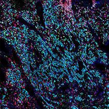
The Cellular Intricacies of the Human Placenta
Ida Emilie Steinmark, PhD | Jul 5, 2023 | 2 min read
Rare samples saved 35 years ago helped researchers map gene expression and cell differentiation in first trimester placentas.
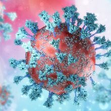
An Introduction to Glycoproteins
Rebecca Roberts, PhD | 5 min read
Making up the majority of all eukaryotic proteins, glycoproteins have a wide range of important physiological roles, from cell-cell signaling to disease pathogenesis.
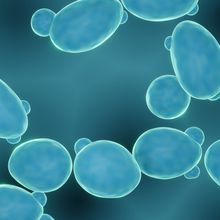
Yeast Cells Reconfigure Their Metabolomes to Live Longer
Danielle Gerhard, PhD | Jul 5, 2023 | 2 min read
Yeast cells share metabolites through extracellular space to extend the lifespan of the entire community.
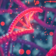
Redesigning Medicine Using Synthetic Biology
Alison Halliday, PhD, Technology Networks | Jun 21, 2023 | 5 min read
Drawing inspiration from nature, synthetic biology offers exciting opportunities to transform the future of medicine.
Automating Circulating Tumor Cell Isolation and Transfer
The Scientist’s Creative Services Team | 1 min read
Darius Cameron Wilson will discuss how the latest automated cell isolation technology streamlines circulating tumor cell applications.
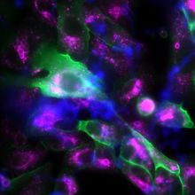
Fungal Spores Hijack a Host Protein to Escape Death
Mariella Bodemeier Loayza Careaga, PhD | Jun 20, 2023 | 3 min read
Uncovering the components used by Aspergillus fumigatus to avoid intracellular destruction broadens our understanding of the mold’s pathogenesis.
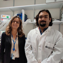
Microglia Rescue Aggregate-Burdened Neurons
Charlene Lancaster, PhD | Jun 12, 2023 | 4 min read
Researchers discover that neurons trade protein aggregates for microglial-derived mitochondria through tunneling nanotubes.
Science Summarized: Smart Labs Connect Scientists, Instruments, and Data
The Scientist’s Creative Services Team and MilliporeSigma | 1 min read
Smart laboratory instruments are intuitively-designed ergonomic tools that support greater connectivity and productivity.
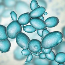
Waves of Macromolecule Production During the Cell Cycle
Mariella Bodemeier Loayza Careaga, PhD | Jun 1, 2023 | 3 min read
In individual yeast cells, essential biosynthetic processes peak at different times in the cell cycle, revealing a temporal dynamic once thought limited to DNA synthesis.

Garbage to Guts: The Slow-Churn of
Plastic Waste
Iris Kulbatski, PhD | Jun 1, 2023 | 4 min read
The winding trail of environmental microplastics leads researchers to the human digestive ecosystem.
Best Practices for Organoid Technologies
The Scientist’s Creative Services Team | 1 min read
Dosh Whye will discuss best practices for organoid modeling and how researchers leverage the latest technologies to achieve their goals.
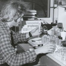
Cellular Competence: Making Recombinant DNA Accessible
Nathan Ni, PhD | Jun 1, 2023 | 2 min read
Coaxing bacteria into taking up recombinant DNA was arduous until Douglas Hanahan took action.
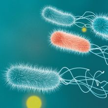
Cooperation and Cheating
Mariella Bodemeier Loayza Careaga, PhD | Jun 1, 2023 | 6 min read
Bacteria cooperate to benefit the collective, but cheaters can rig the system. How is the balance maintained?
