Login
Subscribeimaging
Short-lived Molecules Support Long-term Memory
Alejandra Manjarrez, PhD | Jun 6, 2023 | 3 min read
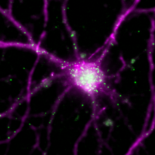
A gene essential for information storage in the brain engages an autoregulatory feedback loop to consolidate memory.
2022 Top 10 Innovations
The Scientist Staff | Dec 12, 2022 | 10+ min read
This year’s crop of winning products features many with a clinical focus and others that represent significant advances in sequencing, single-cell analysis, and more.
See Beyond the Scatter Plot with Imaging, Spectral Flow Cytometry
The Scientist’s Creative Services Team and BD Biosciences | 3 min read
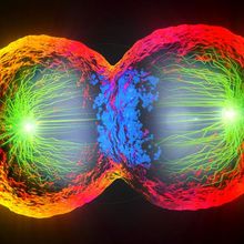
A novel instrument combines fluorescence-activated cell sorting, imaging flow cytometry, and spectral flow cytometry to advance cell population examination.

Obstetrics “Giant” Beryl Benacerraf Dies at 73
Katherine Irving | Oct 26, 2022 | 2 min read
Benacerraf pioneered the use of ultrasound to diagnose fetal syndromes.
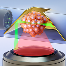
Infographic: Generating Hundreds of 3D Organoid Images per Hour
Natalia Mesa, PhD | Oct 17, 2022 | 1 min read
By modifying a technique used to image single cells, researchers have managed to generate a super-resolution 3D image of a complete organoid in just seven seconds.

Science Philosophy in a Flash - A Look at Aging Through Young Eyes
Iris Kulbatski, PhD | 1 min read
Aimée Parker shares how her childlike curiosity and collaborative spirit motivate her scientific pursuits.

Expert JeWell-ry Designers
Natalia Mesa, PhD | Oct 17, 2022 | 3 min read
Analyzing organoids has proven slow and cumbersome for scientists. But a new technique may speed things up, producing 3D images of hundreds of organoids per hour.
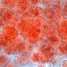
Mouse Brains Appear to Eavesdrop on Their Fat
Alejandra Manjarrez, PhD | Sep 9, 2022 | 4 min read
For the first time, a team visualizes sensory nerves projecting into adipose tissue in mice and finds these neuronal cells may counteract the local effects of the sympathetic nervous system.
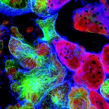
Seeing is Believing with Automated Single Cell Precision Imaging
The Scientist’s Creative Services Team and Canopy Biosciences | 1 min read
Through the Looking Glass: Aging, Inflammation, and Gut Rejuvenation
Iris Kulbatski, PhD | Aug 8, 2022 | 4 min read
Renewing the aging gut microbiome holds promise for preventing inflammatory brain and eye degeneration.
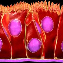
Move Over Apoptosis: Another Form of Cell Death May Occur in the Gut
Natalia Mesa, PhD | May 18, 2022 | 6 min read
Though scientists don’t yet know much about it, a newly described process called erebosis might have profound implications for how the gut maintains itself.
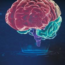
Mapping the Brain in 3-D
Nathan Ni, PhD | 1 min read
3-D brain atlases help scientists better understand brain function in physiological and pathological situations.
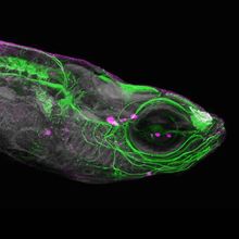
Caught on Camera
The Scientist Staff | May 16, 2022 | 2 min read
See some of the coolest images recently featured by The Scientist
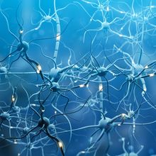
CRACK Method Reveals Novel Neuron Type in Mouse Brain
Dan Robitzski | Apr 18, 2022 | 3 min read
A new technique reveals cells’ precise locations and functions in the brain. Its developers have already used it to identify a previously unknown neuron type.

Demystifying the Black Box
The Scientist’s Creative Services Team and Tecan | 1 min read
One sophisticated laboratory tool detects absorbance, fluorescence, FRET, luminescence, live cells, and more, facilitating a wide variety of experimental findings.
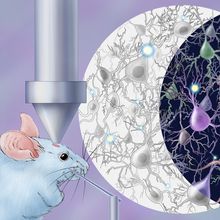
Infographic: Simultaneously Studying Neuron Structure and Function
Dan Robitzski | Apr 18, 2022 | 1 min read
A new methodology combines existing techniques to reveal the specific function and location of multiple types of neurons at once.

2021 Top 10 Innovations
The Scientist Staff | Dec 1, 2021 | 10+ min read
The COVID-19 pandemic is still with us. Biomedical innovation has rallied to address that pressing concern while continuing to tackle broader research challenges.

Form Determines Function: Insights from Structural Biology
The Scientist’s Creative Services Team | 1 min read
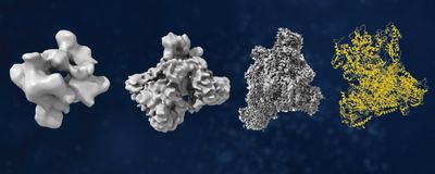
Researchers use diverse tools to analyze protein structures.
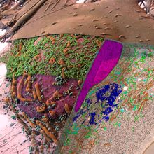
New Studies Enable a Clearer View Inside Cells
Andrew Chapman | Nov 4, 2021 | 5 min read
Armed with improved imaging techniques and supercomputers, researchers are generating detailed three-dimensional images of cellular structures that anyone can explore.
